Upper Inner Thigh Anatomy - inner thigh pain - 630 anatomical structures of the upper limb (pectoral girdle, shoulder, arm, elbow, forearm, wrist, hand and fingers) were labeled.
byAdmin•
0
Upper Inner Thigh Anatomy - inner thigh pain - 630 anatomical structures of the upper limb (pectoral girdle, shoulder, arm, elbow, forearm, wrist, hand and fingers) were labeled.. Gluteus maximus, gluteus medius, gluteus minimus, tensor fasciae latae inner hip muscles. They attach on the inside of your femur ( thighbone ) and on the other end on the pubic bone and sitbones. Upper thigh anatomy (page 1). Vascular anatomy of the upper arm. Inner thigh fat is usually softer and less fibrous than fat found in other areas of the body.
This small fat compartment located in the upper thigh is responsible for excessive friction when you walk, run, or engage in other activities. The single bone in the thigh is called the femur. Want to learn more about it? Of inner thigh lift using a novel dermoadipose ß ap design, which allows an effective anchoring of the inferior ß ap of the. These nerves give sensation to our upper.
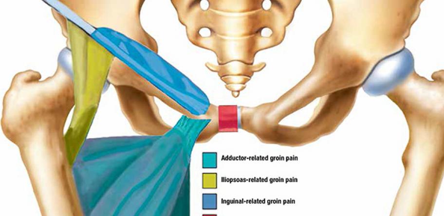
Case Report: Getting to grips with groin pain | How to Treat from adg-qa-assets.s3.ap-southeast-2.amazonaws.com Of inner thigh lift using a novel dermoadipose ß ap design, which allows an effective anchoring of the inferior ß ap of the. The adductor group, commonly known as the inner ( upper) thighs, is a group of several muscles that, when engaged, move the legs together. Palmar region , arteries (illustrations: 935 x 1601 jpeg 153 кб. The information contained in anatomy atlases is not a substitute for the medical care and advice of your physician. Gluteal tuberosity and upper 1/4 of linea aspera. The videos in this series are educational. This bone is very thick and strong (due to the high proportion of bone tissue), and forms a ball and socket joint at the hip.
This inner thigh exercise provides a unique training stimulus for the lower body that will shape your glutes the inner thigh exercise is small but powerful.
Muscle anatomy inner thigh, muscle anatomy of thigh, muscle anatomy of upper thigh, muscle anatomy posterior thigh, muscle anatomy thigh mri, human muscles, muscle anatomy inner thigh, muscle anatomy of thigh, muscle anatomy of upper thigh, muscle anatomy posterior thigh. These pictures of this page are about:upper thigh anatomy. Sprinkle in these inner thigh exercises throughout your exercise routine, or end your workout with a quick inner thigh circuit. They consist of 5 muscles; This bone is very thick and strong (due to the high proportion of bone tissue), and forms a ball and socket joint at the hip. After anatomical analysis of the medial thigh, noting skin and fat redundancy, patients were selected for either an upper/inner medial thighplasty or an extended medial thighplasty. These images are a random sampling from a bing search on the term thigh anatomy. Gluteal tuberosity and upper 1/4 of linea aspera. The information contained in anatomy atlases is not a substitute for the medical care and advice of your physician. The muscles, bones, joints, nerves, blood and lymphatic supply, anatomical areas, and the the nerves of the upper limb arise from a complex arrangement of nerve fibers known as the brachial plexus; Linea aspera and popliteal surface minimus: Thigh anatomy is fairly complex, since we are talking about two of the main joints in the human body: This inner thigh exercise provides a unique training stimulus for the lower body that will shape your glutes the inner thigh exercise is small but powerful.
The upper leg muscles stock illustration. You should feel the inner upper thigh tone or activate slightly. These images are a random sampling from a bing search on the term thigh anatomy. Linea aspera and popliteal surface minimus: This webpage presents the anatomical structures found on thigh mri.
Leg artery anatomy. Cardiovascular System of the Leg and ... from mrimaster.com They attach on the inside of your femur ( thighbone ) and on the other end on the pubic bone and sitbones. These nerves give sensation to our upper. Rectus femoris, vastus lateralis, vastus medialis, and vastus intermedius. Vascular anatomy of the upper arm. Gluteus maximus, gluteus medius, gluteus minimus, tensor fasciae latae inner hip muscles. There are 7 main areas covered in the upper limb; Thigh anatomy is fairly complex, since we are talking about two of the main joints in the human body: Gluteal tuberosity and upper 1/4 of linea aspera.
They consist of 5 muscles;
Learn vocabulary, terms and more with flashcards, games and other study tools. 370 x 415 gif 19 кб. Anatomically, it is part of the lower limb. These images are a random sampling from a bing search on the term thigh anatomy. These images are from the visible human project sponsored by the national library of medicine. Gluteal tuberosity and upper 1/4 of linea aspera. Vascular anatomy of the upper arm. There may be variations in treatment that. Anatomylearning.com/en/ intro template created by: Learn proper form, discover all health benefits and choose a workout. Gluteus maximus, gluteus medius, gluteus minimus, tensor fasciae latae inner hip muscles. The inner thighs are meant to assist in a internal rotation that helps to activate the psoas muscle. This webpage presents the anatomical structures found on thigh mri.
Appendicular muscles of the pelvic girdle and lower limbs. It is your full responsibility to be cautious when attempting the techniques, stop if it becomes uncomfortable. Gluteus maximus, gluteus medius, gluteus minimus, tensor fasciae latae inner hip muscles. The inner thighs are meant to assist in a internal rotation that helps to activate the psoas muscle. This webpage presents the anatomical structures found on thigh mri.
Muscle and Tendon Characteristics - Classic Human Anatomy ... from doctorlib.info Palmar region , arteries (illustrations: In human anatomy, the thigh is the area between the hip (pelvis) and the knee. Gluteus maximus, gluteus medius, gluteus minimus, tensor fasciae latae inner hip muscles. The exercises for inner thighs mentioned will help you tone up and strengthen the muscles of your inner thigh adductor anatomy: There may be variations in treatment that. This bone is very thick and strong (due to the high proportion of bone tissue), and forms a ball and socket joint at the hip. These images are a random sampling from a bing search on the term thigh anatomy. Vascular anatomy of the upper arm.
This webpage presents the anatomical structures found on thigh mri.
A tutorial covering the position, actions, attachments and innervation of the gracilis muscle with the aid of detailed anatomical illustrations and a quiz. Learn proper form, discover all health benefits and choose a workout. Inner thigh fat is usually softer and less fibrous than fat found in other areas of the body. It is your full responsibility to be cautious when attempting the techniques, stop if it becomes uncomfortable. Learn vocabulary, terms and more with flashcards, games and other study tools. Sometimes the inner thigh muscles (the adductors) can feel really tight, even if you haven't been training. After anatomical analysis of the medial thigh, noting skin and fat redundancy, patients were selected for either an upper/inner medial thighplasty or an extended medial thighplasty. The muscles, bones, joints, nerves, blood and lymphatic supply, anatomical areas, and the the nerves of the upper limb arise from a complex arrangement of nerve fibers known as the brachial plexus; Learn how to use inner thigh exercises each of the muscles in the inner thigh has different actions. This small fat compartment located in the upper thigh is responsible for excessive friction when you walk, run, or engage in other activities. 370 x 415 gif 19 кб. Below, it tapers into a skinny tendon that follows the bulk of sartorius into the medial condyle of the tibia. This webpage presents the anatomical structures found on thigh mri.
Sprinkle in these inner thigh exercises throughout your exercise routine, or end your workout with a quick inner thigh circuit upper thigh anatomy. Inner thigh fat is usually softer and less fibrous than fat found in other areas of the body.